| Home | E-Submission | Sitemap | Contact Us |  
top_img
Radiation Oncology Journal > Volume 37(2); 2019 > Article
Sharma, Ahlawat, Gairola, Tandon, Sachdeva, and Sharief: Prognostic factors, failure patterns and survival analysis in patients with resectable oral squamous cell carcinoma of the tongue

Abstract

Purpose

There is sparse literature on treatment outcomes research on resectable oral tongue squamous cell carcinoma (OTSCC). The aim of this study was to measure the treatment outcomes, explore the failure patterns, and identify the potential clinicopathological prognostic factors affecting treatment outcomes for resectable OTSCC.

Materials and Methods

It is a retrospective analysis of 202 patients with resectable OTSCC who underwent upfront primary surgical resection followed by adjuvant radiotherapy with or without concurrent chemotherapy if indicated.

Results

The median follow-up was 35.2 months (range, 1.2 to 99.9 months). The median duration of locoregional control (LRC) was 84.9 months (95% confidence interval, 67.3–102.4). The 3- and 5-year LRC rate was 68.5% and 58.3%, respectively. Multivariate analysis showed that increasing pT stage, increasing pN stage, and the presence of extracapsular extension (ECE) were significantly associated with poorer LRC. The median duration of overall survival (OS) was not reached at the time of analysis. The 3- and 5-year OS rate was 70.5% and 66.6%, respectively. Multivariate analysis showed that increasing pT stage and the presence of ECE were significantly associated with a poorer OS.

Conclusion

Locoregional failure remains the main cause of treatment failure in resectable OTSCC. There is scope to further improve prognosis considering modest LRC and OS. Pathological T-stage, N-stage, and ECE are strong prognostic factors. Further research is required to confirm whether adjuvant therapy adds to treatment outcomes in cases with lymphovascular invasion, perineural invasion, and depth of invasion, and help clinicians tailoring adjuvant therapy.

Introduction

Incidence of oral tongue squamous cell carcinoma (OTSCC) is on rise especially in the younger population [1-3], probably due to a rise in tobacco and alcohol intake. Early stage OTSCC is treated with single modality therapy preferably surgery [4], whereas locally advanced resectable disease is treated with combined modality therapy—surgery followed by adjuvant therapy with radiotherapy (RT) or chemoradiation (CRT). Although several clinicopathological prognostic factors have been found for oral cavity squamous cell carcinoma (OCSCC), most of these have been reported in studies done on mixed patient population with all subsites of oral cavity combined [5,6]. Data is limited for OTSCC per se. Since OTSCC is on rise and treatment outcome of OTSCC has been found to be poorer than that of carcinoma arising from other subsites of oral cavity [7,8], it is important to identify clinicopathological factors for carcinoma arising from this subsite. The aim of this study was to measure the treatment outcomes, explore the failure patterns, and identify the potential clinicopathological prognostic factors affecting treatment outcomes for resectable OTSCC.

Materials and Methods

The Institutional Ethics Committee of our institution approved the study. The inclusion criteria were patients with localized and resectable OTSCC with or without cervical nodal involvement who had undergone primary surgical resection of the primary tumor and regional lymph nodes at this institute between November 2010 and December 2016. The exclusion criteria included: an Eastern Cooperative Oncology Group performance status of ≥2; neoadjuvant chemotherapy administered prior to primary surgical resection; surgical resection performed with palliative or debulking intent; recurrent head and neck malignancies; and a prior history of RT to the head and neck region. Although the American Joint Committee on Cancer (AJCC) 7th edition was used for the staging and management, AJCC 8th edition was used to analyze prognostic factors in the analysis of this study. Adjuvant RT was administered within 4 to 6 weeks from the date of surgery depending on the high-risk features recorded in the histopathological report of the resected specimen. As per institutional policy, the following were the indications for adjuvant RT: pathological T3 or T4 stage, node positivity (even a single node), positive resection margins (<1.0 mm), close surgical margins (≥1.0 mm to ≤4.0 mm), perineural invasion (PNI), and lymphovascular invasion (LVI). Depth of invasion (DOI) of >4.0 mm was considered as an indication for adjuvant RT depending on the individual policy of treating physicians. Concurrent chemotherapy—weekly cisplatin (40.0 mg/m2) or carboplatin (area under the curve 2) was administered to patients with positive resection margins or extracapsular extension (ECE). Whilst planning adjuvant CRT, patients were initially considered for weekly cisplatin. Those who were elderly or presumed unfit for cisplatin were administered carboplatin or cetuximab.
Patients were treated with two-dimensional conventional RT or simultaneous-integrated boost intensity-modulated RT (SIB-IMRT). Conventional fractionation was used for all patients. For SIB-IMRT surgical bed with positive margin or nodal region with ECE were given 66 Gy. Surgical bed without positive margin or nodal regions without ECE were given 60 Gy in 30 fractions. Elective nodal regions were given 54 Gy in 30 fractions. When two-dimensional parallel opposed shrinking field technique was used we delivered 50 Gy in 25 fractions to elective nodal regions and boosted surgical bed without margin positive or nodal regions without ECE to 10 Gy in 5 fractions. Surgical bed with positive margin or nodal region with ECE was boosted to another 6 Gy in 3 fractions.

1. Statistical analysis

All statistical analyses were performed using statistical package for the social science system (SPSS version 20; IBM SPSS, Armonk, NY, USA), and p-value less than 0.05 was considered statistically significant. All p-values reported represent two-sided tests. Baseline clinical and pathological categorical variables were expressed as frequencies along with respective percentages. The endpoints were locoregional control (LRC) and overall survival (OS), and were calculated by the Kaplan–Meier product-limit method. Univariate analysis of LRC and OS was performed on the following clinical and histopathological factors selected based on results from previous studies on oral cavity cancer: age, sex, addictions (tobacco smoking, tobacco chewing and/or alcohol consumption), tumor grade, pathological T (pT) stage, pathological N (pN) stage, PNI, LVI, resection margin status, DOI, and ECE. Multivariate Cox proportional hazards regression analysis was performed to estimate the impact of known relevant prognostic factors.

Results

The medical records of 202 patients fulfilling inclusion and exclusion criteria were reviewed. Patient and disease-related characteristics are shown in Table 1. The median follow-up was 35.2 months (range, 1.2 to 99.9 months).
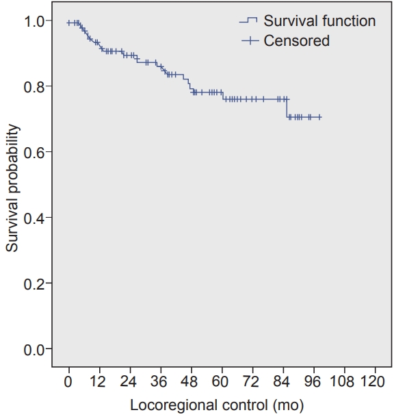
The median duration of LRC was 84.9 months (95% confidence interval, 67.3–102.4). The 3- and 5-year LRC rate was 68.5% and 58.5%, respectively (Fig. 1). Univariate analysis showed that increasing pT stage, increasing pN stage, and the presence of PNI, LVI, and ECE were significant poor prognostic factors for LRC. However, multivariate analysis showed that increasing pT stage, increasing pN stage, and the presence of ECE were significantly associated with poorer LRC (Table 2).
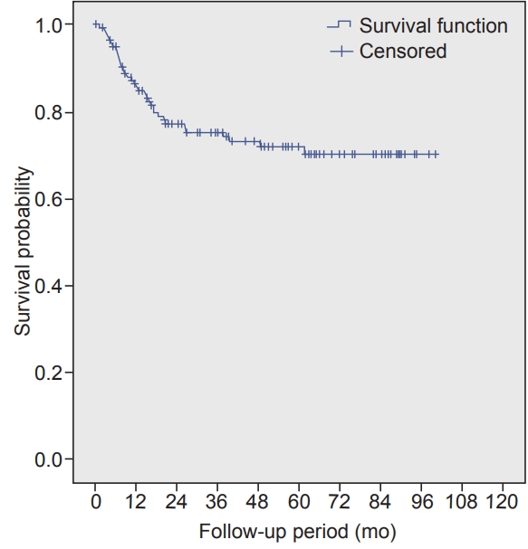
The median duration of OS was not reached at the time of analysis. The 3- and 5-year OS rate was 70.5% and 66.6, respectively (Fig. 2). Univariate analysis revealed that increasing pT stage, increasing pN stage, presence of LVI, DOI of >20 mm, and ECE were poor prognostic factors for OS. However, multivariate analysis using Cox proportional hazard ratios showed that increasing pT stage and the presence of ECE were significantly associated with a poorer OS (Table 3).
Majority of percent patients completed planned treatment. At the time of analysis, 74 patients (36.6%) had developed recurrence. Of these, 33 patients (44.5%) experienced tumor recurrence in the primary site alone, 23 (31.1%) experienced recurrence in the nodal region alone, 3 (4.0%) experienced recurrence in the primary site and nodal region, and 15 (20.2%) had distant metastases most common being lung followed by bone.

Discussion and Conclusion

This study is an attempt to determine LRC and OS, failure pattern and explore various prognostic clinical and pathological factors influencing LRC and OS for OTSCC. This study continued to provide yet another evidence that locoregional recurrence remains a major mode of treatment failure in this subset of patients, considering approximately a third of recurrence being locoregional in this study. Hence, thorough attempt should be made to identify high-risk patients for recurrence based on clinical and pathological factors for whom additional therapy in the form of adjuvant RT or CRT may improve outcomes.
Although there were 38.6% patients in the current study who were found to have pathological stage III/IV, more than two-thirds of patients from the entire cohort received adjuvant RT because of combination of other risk factors on surgical histopathological specimen examination. Approximately one-fourth amongst those who received adjuvant RT also received concurrent chemotherapy (CCT).
Gender may have an influence on the treatment outcome as shown by various studies with large sample population including all subsites of oral cavity combined together [9-12]. These studies showed female gender being associated with better survival. The current study did not show any association with outcomes in line with Garavello et al. [13] who particularly looked at the influence of gender on OTSCC similar to our study, and found that gender does not influence prognosis in this subsite. Age whether as continuous or categorical variable was not found to affect outcome in OTSCC in this study. However in a Surveillance, Epidemiology and End Results database analysis, there was found to be a significant difference in median OS between age groups: 50–69 years and ≥70 years with OTSCC (59.5 vs. 73.1 months, respectively); however the difference nullified when results were analysed after stage stratification [14]. Elderly patients tend to have multiple comorbidities which may influence the choice of therapy, intent of therapy, course of intensive treatment such as major surgeries or adjuvant CRT and may have poor tolerance to treatment, thereby showing different response to therapy. Thus, it appears that association of prognosis with age is complex and less clearly understood as yet in OTSCC.
A number of reports have showed that alcohol consumption and smoking are prognostic factor for head and neck cancer [15-18]. When looking at OTSCC per se, Sawabe et al. [19] in a prospective study showed that patients with OTSCC treated with surgery is associated with a significant inverse association between alcohol consumption and prognosis. However we did not find these habits be a prognostic factor in Indian population particularly looking at OTSCC. Our results are similar to that of a study done by Thiagarajan et al. [20] on similar cohort of patients as ours (Indian patients with OTSCC), that habits (including alcohol, smoking and tobacco chewing) does not affect the OS.
In the OTSCC, T-stage and N-stage are independent indicators of the prognosis, although they are inter-related. Increasing size by T-stage leads to an increase in the rate of occult metastases [21-23], and increasing N-stage is associated with the development of distant metastases, particularly with multiple involvement or the presence of ECE [22]. Increasing pT stage emerged from our study as being of high prognostic value, as the risk of local recurrence at the time of diagnosis increased when tumour size increases from less than 2 cm to more than 4 cm. The benefit in OS increased as the pT stage increased from T2 to T4. This experience is similar to results found in other reports [24].
Presence of cervical lymphadenopathy is shown as single most important clinical predictor of OS in this study. However, mere presence of single node less than 3 cm did not affect OS, it was the presence of multiple nodes which affected OS significantly poorly. There were no patients with clinical N3 stage because surgeons found them to be unresectable, and hence were given radical RT/CRT. There were no patients who were upstaged to pN3 stage either. All patients received CCT in this study if there were ECE. This was based on the combined analysis of landmarks trials EORTC-22931 and RTOG-9501 studies which showed advantage of adding CCT [25]. Despite adding CCT in the presence of ECE in our study patients, it still continued to show significant impact on OS on multivariate analysis. When looking particularly at OTSCC high quality data is sparse regarding the impact of ECE. Our results are consistent with few retrospective studies which showed ECE as a significant predictor of OS in OTSCC [20,26,27].
DOI is used as a surrogate for risk of lymphatic involvement. Hence, neck dissection (elective neck dissection) has become an acceptable neck management strategy for OTSCC when the DOI is high [28,29]. Most centers regard 3.0 to 4.0 mm as the optimal cutoff for DOI, beyond which neck management is essential for the clinically node-negative neck. There has been considerable controversy regarding the need for adjuvant RT in patients without high-risk features, except for a DOI of >4.0 mm. Following the results published by Ganly et al. [30], from the Memorial Sloan Kettering Cancer Center and Princess Margaret Hospital in 2013, which demonstrated much higher regional failure than anticipated in patients with pT1/2 and pN0 OSCC, who underwent partial glossectomy and ipsilateral elective neck dissection without postoperative RT, our institution has accepted a DOI of >4.0 mm alone as a sufficient risk factor to offer adjuvant RT in the majority of these cases, although it influenced OS in this study in univariate analysis only when it was >20 mm. Few other studies have used multivariate analysis to substantiate the importance of the DOI in OTSCC or have revealed a correlation between the DOI and LRC or disease-free survival [31-33]. A recent study by Gokavarapu et al. [34] showed that postoperative RT did not influence LRC or OS in a subset of patients with pT1/2 and pN0 OSCC with a DOI of ≥4.0 mm. Although it appears certain that DOI has prognostic role for OCSCC, it is yet to be evaluated beyond which DOI in mm adjuvant RT benefit significantly.
LVI has been shown to be significantly associated with tumor site, diameter, and thickness, PNI, the invasive front, the pattern of invasion, lymph node metastasis, resection margin status, local recurrence, and survival [22,35-37]. Consistent with these findings, our study showed a significant association between LVI and OS in the univariate analysis. However, in the multivariate analysis, LVI was not found to be an independent predictor of OS. A review of the literature identified only one study that reported LVI to be significant in the multivariate analysis. Rahima et al. [38] discussed the possibility of using this parameter as a criterion to define aggressiveness and to select patients for more specific and aggressive treatment in the future.
In the analysis by El-Husseiny et al. [39] on OTSCC (T1-4 N0-3) the reported 5-year OS was 65%. Rusthoven et al. [7] described a 5-year OS of 60.9% in patients treated for T1-2 N0 OTSCC. Although the prognosis of OTSCC was poorer than that of cancers in other oral cavity sites, in this study, the 5-year OS and LRC were shown as 66.6% and 58.3%, respectively. This study showed the survival outcome of OTSCC to be affected by increasing T stage, more than two clinically positive nodes, ECE of lymph node metastasis and LVI on univariate analysis. These findings have also been reported by others [24,40-42]. However, multivariate analysis revealed increasing pT stage and presence of ECE to be significantly associated with overall survival.
The limitations of this study include potential bias associated with its retrospective design. Although the treatment guidelines followed at our institute are standardized, some differences remain in the indications of adjuvant therapy in OCSCC depending on individual experiences of the treating oncologist, especially with regards to DOI. A strength of this study is its large cohort of patients with squamous cell carcinoma limited to oral tongue subsite only, taken from one of the largest and most exclusive cancer research centers in the country. Furthermore, the fact that all surgical and histopathological reporting was done at a single institute may have helped to reduce bias.
In conclusion, locoregional failure remains the main cause of treatment failure in resectable OTSCC. There is scope to further improve prognosis considering modest LRC and OS. Pathological T-stage, N-stage, and ECE are strong prognostic factors. Further research is required to confirm whether adjuvant therapy adds to treatment outcomes in cases with LVI, PNI and DOI, and help clinicians tailoring adjuvant therapy.

Notes

Conflict of Interest

No potential conflict of interest relevant to this article was reported.

Fig. 1.
Kaplan-Meier curve for locoregional control.
roj-2018-00577f1.jpg
Fig. 2.
Kaplan-Meier curve for overall survival.
roj-2018-00577f2.jpg
Table 1.
Patient and disease-related characteristics
Variable Value
Age (yr) 54.19 ± 14.16
 Median (range) 56 (23–94)
 ≤50 81 (40.1)
 >50 121 (59.9)
Sex
 Male 153 (75.7)
 Female 49 (24.3)
Addictions
 Absent 97 (48.1)
 Tobacco 86 (81.9)
 Alcohol 32 (30.5)
Grade
 Well-differentiated 49 (24.3)
 Moderately differentiated 148 (73.3)
 Poorly differentiated 5 (2.4)
Type of surgery
 Wide local excision 32 (15.8)
 Hemiglossectomy 170 (84.2)
Type of neck dissection
 Selective nodal dissection 133 (65.8)
 Modified neck dissection 43 (21.3)
 Radical neck dissection 19 (9.4)
 Not dissected 7 (3.5)
pT Stage
 pT1 37 (18.3)
 pT2 101 (50.0)
 pT3 51 (25.3)
 pT4a 13 (6.4)
pN Stage
 pN0 123 (60.2)
 pN1 25 (12.8)
 pN2a 10 (5.0)
 pN2b 9 (4.5)
 pN2c 3 (1.5)
 pN3b 25 (12.5)
 Not dissected 7 (3.5)
Stage
 I 30 (14.9)
 II 65 (32.2)
 III 48 (23.7)
 IVa 27 (13.4)
 IVb 25 (12.3)
 Unknown 7 (3.5)
Adjuvant radiotherapy
 Received 144 (71.3)
 Non received 58 (29.7)
Type of radiotherapy
 Conventional 36/144 (25)
 IMRT 108/144 (75)
Concurrent chemotherapy
 Received 38/144 (26.4)
 Non received 106/144 (73.6)
High-risk features
 pT3/4 27 (13.4)
 pN+ve 72 (36.9)
 ECE, present 31/72 (43.1)
 Margins, positive 15 (7.4)
 Margins, close 50 (24.7)
 LVI, present 53 (26.2)
 PNI, present 76 (37.6)
 DOI (mm)
  ≤5 64 (31.7)
  6–10 88 (43.6)
  >10 and ≤20 46 (22.7)
  >20 4 (2.0)

Values are presented as mean ± standard deviation or number (%).

ECE, extracapsular extension; LVI, lymphovascular invasion; PNI, perineural invasion; DOI, depth of invasion; IMRT, intensity-modulated radiotherapy.

Table 2.
Univariate and multivariate analysis for locoregional control
Variable Univariate
Multivariate
HR (95% CI) p-value HR (95% CI) p-value
Age (yr) 1.652 (0.476–2.067) 0.415
 >50 Ref
 ≤50 0.603 (0.466–1.209) 0.771
Sex
 Male Ref
 Female 1.219 (0.477–2.190) 0.702
Addictions
 No Ref
 Yes 0.606 (0.398–2.320) 0.828
Grade
 Well-differentiated Ref
 Moderately differentiated 1.549 (0.475–2.832) 0.493
 Poorly differentiated 4.128 (0.757–13.012) 0.098
pT Stage
 pT1 Ref Ref
 pT2 2.043 (0.956–4.369) 0.055 1.967 (0.901–4.291) 0.089
 pT3 2.525 (1.113–5.731) 0.027 2.453 (1.033–5.824) 0.042
 pT4 6.815 (2.184–21.269) 0.001 5.029 (1.207–20.950) 0.027
pN Stage
 pN0 Ref Ref
 pN1 0.643 (0.290–1.427) 0.207 0.494 (0.213–1.146) 0.100
 pN2 1.303 (0.656–2.589) 0.450 1.632 (0.378–2.204) 0.839
 pN3 2.417 (1.237–4.723) 0.010 2.166 (1.376–8.141) 0.039
Margin
 Adequate Ref
 Close 2.073 (0.743–3.630) 0.642
 Positive 1.092 (0.332–3.882) 0.157
ECE
 Absent Ref Ref
 Present 5.773 (3.091–8.653) 0.005 3.086 (1.268–4.400) 0.018
LVI
 Absent Ref Ref
 Present 3.033 (1.112–5.533) 0.017 1.274 (0.725–2.240) 0.400
PNI
 Absent Ref Ref
 Present 1.982 (0.344–4.036) 0.021 1.504 (0.543–1.904) 0.197
DOI (mm)
 ≤5 Ref
 6–10 1.192 (0.678–2.097) 0.542
 >10 and ≤20 1.427 (0.747–2.729) 1.427
 >20 3.826 (0.492–29.751) 0.200
Type of radiotherapy
 IMRT Ref
 Conventional 0.623 (0.387–3.77) 0.766

HR, hazard ratio; CI, confidence interval; ECE, extracapsular extension; LVI, lymphovascular invasion; PNI, perineural invasion; DOI, depth of invasion; IMRT, intensity-modulated radiotherapy.

Table 3.
Univariate and multivariate analysis for overall survival
Variable Univariate
Multivariate
HR (95% CI) p-value HR (95% CI) p-value
Age (yr) 1.328 (0.768–2.297) 0.310
 >50 Ref
 ≤50 0.997 (0.977–1.018) 0.805
Sex
 Male Ref
 Female 1.007 (0.546–1.855) 0.983
Addictions
 No Ref
 Yes 0.918 (0.536–1.573) 0.755
Grade
 Well-differentiated Ref
 Moderately differentiated 1.288 (0.659–2.517) 0.460
 Poorly differentiated 3.242 (0.898–11.706) 0.073
pT Stage
 pT1 Ref Ref
 pT2 5.467 (1.302–22.956) 0.020 6.061 (1.294–28.385) 0.022
 pT3 7.605 (1.762–32.815) 0.007 14.876 (2.668–82.945) 0.002
 pT4 25.505 (5.062–128.510) 0.000 16.809 (2.053–137.644) 0.009
pN Stage
 pN0 Ref Ref
 pN1 1.650 (0.731–3.725) 0.228 1.025 (0.420–2.499) 1.025
 pN2 2.308 (1.021–5.215) 0.044 1.184 (0.426–3.292) 0.747
 pN3 6.942 (3.627–13.288) 0.000 4.268 (0.873–27.047) 0.070
Margin
 Adequate Ref
 Close 1.095 (0.592–2.026) 0.772
 Positive 1.725 (0.592–2.026) 0.258
ECE
 Absent Ref Ref
 Present 4.154 (2.359–7.314) 0.000 3.085 (1.214–5.498) 0.028
LVI
 Absent Ref Ref
 Present 2.392 (1.393–4.107) 0.002 1.665 (0.911–3.044) 0.098
PNI
 Absent Ref
 Present 1.604 (0.930–2.769) 0.090
DOI (mm)
 ≤5 Ref Ref
 6–10 1.585 (0.799–3.143) 0.187 0.872 (0.404–1.880) 0.726
 >10 and ≤20 1.455 (0.653–3.243) 0.359 1.285 (0.096–2.839) 0.323
 >20 11.045 (2.298–53.073) 0.030 2.013 (0.253–15.992) 0.508
Type of radiotherapy
 IMRT Ref
 Conventional 0.991 (0.481–2.040) 0.980

HR, hazard ratio; CI, confidence interval; ECE, extracapsular extension; LVI, lymphovascular invasion; PNI, perineural invasion; DOI, depth of invasion; IMRT, intensity-modulated radiotherapy.

References

1. Myers JN, Elkins T, Roberts D, Byers RM. Squamous cell carcinoma of the tongue in young adults: increasing incidence and factors that predict treatment outcomes. Otolaryngol Head Neck Surg 2000;122:44–51.
crossref pmid
2. Annertz K, Anderson H, Biorklund A, et al. Incidence and survival of squamous cell carcinoma of the tongue in Scandinavia, with special reference to young adults. Int J Cancer 2002;101:95–9.
crossref pmid
3. Shiboski CH, Schmidt BL, Jordan RC. Tongue and tonsil carcinoma: increasing trends in the U.S. population ages 20-44 years. Cancer 2005;103:1843–9.
crossref pmid
4. Fein DA, Mendenhall WM, Parsons JT, et al. Carcinoma of the oral tongue: a comparison of results and complications of treatment with radiotherapy and/or surgery. Head Neck 1994;16:358–65.
crossref pmid
5. Lefebvre JL, Coche-Dequeant B, Buisset E, Mirabel X, Van JT, Prevost B. Management of early oral cavity cancer: experience of Centre Oscar Lambret. Eur J Cancer B Oral Oncol 1994;30B:216–20.
crossref pmid
6. Lapeyre M, Bollet MA, Racadot S, et al. Postoperative brachytherapy alone and combined postoperative radiotherapy and brachytherapy boost for squamous cell carcinoma of the oral cavity, with positive or close margins. Head Neck 2004;26:216–23.
crossref pmid
7. Rusthoven K, Ballonoff A, Raben D, Chen C. Poor prognosis in patients with stage I and II oral tongue squamous cell carcinoma. Cancer 2008;112:345–51.
crossref pmid
8. Cheng YJ, Tsai MH, Chiang CJ, et al. Adjuvant radiotherapy after curative surgery for oral cavity squamous cell carcinoma and treatment effect of timing and duration on outcome-A Taiwan Cancer Registry national database analysis. Cancer Med 2018;Jun 14 [Epub]. http://doi.org/10.1002/cam4.1611.
crossref
9. Micheli A, Mariotto A, Giorgi Rossi A, Gatta G, Muti P. The prognostic role of gender in survival of adult cancer patients. EUROCARE Working Group. Eur J Cancer 1998;34(14 Spec No):2271–8.
crossref pmid
10. Funk GF, Karnell LH, Robinson RA, Zhen WK, Trask DK, Hoffman HT. Presentation, treatment, and outcome of oral cavity cancer: a National Cancer Data Base report. Head Neck 2002;24:165–80.
crossref pmid
11. Franco EL, Dib LL, Pinto DS, Lombardo V, Contesini H. Race and gender influences on the survival of patients with mouth cancer. J Clin Epidemiol 1993;46:37–46.
crossref pmid
12. Listl S, Jansen L, Stenzinger A, et al. Survival of patients with oral cavity cancer in Germany. PLoS One 2013;8:e53415.
crossref pmid pmc
13. Garavello W, Spreafico R, Somigliana E, Gaini L, Pignataro L, Gaini RM. Prognostic influence of gender in patients with oral tongue cancer. Otolaryngol Head Neck Surg 2008;138:768–71.
crossref pmid pmc
14. Bhattacharyya N. A matched survival analysis for squamous cell carcinoma of the head and neck in the elderly. Laryngoscope 2003;113:368–72.
crossref pmid
15. Leoncini E, Vukovic V, Cadoni G, et al. Clinical features and prognostic factors in patients with head and neck cancer: results from a multicentric study. Cancer Epidemiol 2015;39:367–74.
crossref pmid
16. Li Y, Mao Y, Zhang Y, et al. Alcohol drinking and upper aerodigestive tract cancer mortality: a systematic review and meta-analysis. Oral Oncol 2014;50:269–75.
crossref pmid
17. Inoue M, Nagata C, Tsuji I, et al. Impact of alcohol intake on total mortality and mortality from major causes in Japan: a pooled analysis of six large-scale cohort studies. J Epidemiol Community Health 2012;66:448–56.
crossref pmid
18. Kim MK, Ko MJ, Han JT. Alcohol consumption and mortality from all-cause and cancers among 1.34 million Koreans: the results from the Korea national health insurance corporation's health examinee cohort in 2000. Cancer Causes Control 2010;21:2295–302.
crossref pmid pdf
19. Sawabe M, Ito H, Oze I, et al. Heterogeneous impact of alcohol consumption according to treatment method on survival in head and neck cancer: a prospective study. Cancer Sci 2017;108:91–100.
crossref pmid pmc
20. Thiagarajan S, Nair S, Nair D, et al. Predictors of prognosis for squamous cell carcinoma of oral tongue. J Surg Oncol 2014;109:639–44.
crossref pmid
21. Kirita T, Ohgi K, Shimooka H, et al. Preoperative concurrent chemoradiotherapy plus radical surgery for advanced squamous cell carcinoma of the oral cavity: an analysis of long-term results. Oral Oncol 1999;35:597–606.
crossref pmid
22. Jones HB, Sykes A, Bayman N, et al. The impact of lymphovascular invasion on survival in oral carcinoma. Oral Oncol 2009;45:10–5.
crossref pmid
23. Yanamoto S, Yamada S, Takahashi H, et al. Clinicopathological risk factors for local recurrence in oral squamous cell carcinoma. Int J Oral Maxillofac Surg 2012;41:1195–200.
crossref pmid
24. Lin CY, Wang HM, Kang CJ, et al. Primary tumor site as a predictor of treatment outcome for definitive radiotherapy of advanced-stage oral cavity cancers. Int J Radiat Oncol Biol Phys 2010;78:1011–9.
crossref pmid
25. Bernier J, Cooper JS, Pajak TF, et al. Defining risk levels in locally advanced head and neck cancers: a comparative analysis of concurrent postoperative radiation plus chemotherapy trials of the EORTC (#22931) and RTOG (#9501). Head Neck 2005;27:843–50.
crossref pmid
26. Myers JN, Greenberg JS, Mo V, Roberts D. Extracapsular spread: a significant predictor of treatment failure in patients with squamous cell carcinoma of the tongue. Cancer 2001;92:3030–6.
crossref pmid
27. Metcalfe E, Aspin L, Speight R, et al. Postoperative (chemo) radiotherapy for oral cavity squamous cell carcinomas: outcomes and patterns of failure. Clin Oncol (R Coll Radiol) 2017;29:51–9.
crossref pmid
28. Fukano H, Matsuura H, Hasegawa Y, Nakamura S. Depth of invasion as a predictive factor for cervical lymph node metastasis in tongue carcinoma. Head Neck 1997;19:205–10.
crossref pmid
29. Al-Rajhi N, Khafaga Y, El-Husseiny J, et al. Early stage carcinoma of oral tongue: prognostic factors for local control and survival. Oral Oncol 2000;36:508–14.
crossref pmid
30. Ganly I, Goldstein D, Carlson DL, et al. Long-term regional control and survival in patients with "low-risk," early stage oral tongue cancer managed by partial glossectomy and neck dissection without postoperative radiation: the importance of tumor thickness. Cancer 2013;119:1168–76.
crossref pmid
31. O-charoenrat P, Pillai G, Patel S, et al. Tumour thickness predicts cervical nodal metastases and survival in early oral tongue cancer. Oral Oncol 2003;39:386–90.
crossref pmid
32. Spiro RH, Huvos AG, Wong GY, Spiro JD, Gnecco CA, Strong EW. Predictive value of tumor thickness in squamous carcinoma confined to the tongue and floor of the mouth. Am J Surg 1986;152:345–50.
crossref pmid
33. Larsen SR, Johansen J, Sorensen JA, Krogdahl A. The prognostic significance of histological features in oral squamous cell carcinoma. J Oral Pathol Med 2009;38:657–62.
crossref pmid
34. Gokavarapu S, Parvataneni N, Rao S LM, Reddy R, Raju KV, Chander R. Role of postoperative radiation therapy (PORT) in pT1-T2 N0 deep tongue cancers. Oral Surg Oral Med Oral Pathol Oral Radiol 2015;120:e227–31.
crossref pmid
35. Sparano A, Weinstein G, Chalian A, Yodul M, Weber R. Multivariate predictors of occult neck metastasis in early oral tongue cancer. Otolaryngol Head Neck Surg 2004;131:472–6.
crossref pmid
36. Binmadi NO, Basile JR. Perineural invasion in oral squamous cell carcinoma: a discussion of significance and review of the literature. Oral Oncol 2011;47:1005–10.
crossref pmid
37. Fagan JJ, Collins B, Barnes L, D'Amico F, Myers EN, Johnson JT. Perineural invasion in squamous cell carcinoma of the head and neck. Arch Otolaryngol Head Neck Surg 1998;124:637–40.
crossref pmid
38. Rahima B, Shingaki S, Nagata M, Saito C. Prognostic significance of perineural invasion in oral and oropharyngeal carcinoma. Oral Surg Oral Med Oral Pathol Oral Radiol Endod 2004;97:423–31.
crossref pmid
39. El-Husseiny G, Kandil A, Jamshed A, et al. Squamous cell carcinoma of the oral tongue: an analysis of prognostic factors. Br J Oral Maxillofac Surg 2000;38:193–9.
crossref pmid
40. Kalnins IK, Leonard AG, Sako K, Razack MS, Shedd DP. Correlation between prognosis and degree of lymph node involvement in carcinoma of the oral cavity. Am J Surg 1977;134:450–4.
crossref pmid
41. Woolgar JA, Rogers SN, Lowe D, Brown JS, Vaughan ED. Cervical lymph node metastasis in oral cancer: the importance of even microscopic extracapsular spread. Oral Oncol 2003;39:130–7.
crossref pmid
42. Grandi C, Alloisio M, Moglia D, et al. Prognostic significance of lymphatic spread in head and neck carcinomas: therapeutic implications. Head Neck Surg 1985;8:67–73.
crossref pmid
Editorial Office
Department of Radiation Oncology, Samsung Medical Center,
Proton Therapy Center, B2, 81, Irwon-ro, Gangnam-gu, Seoul 06351, Republic of Korea
Tel : +82-2-3410-3617
E-mail: rojeditor@gmail.com, roj@kosro.or.kr
Copyright © The Korean Society for Radiation Oncology.                      Developed in M2PI
Close layer
prev next